Chapter 8: Cell Division
Cell Division Introduction and Overview
Cell Division
Organisms grow, repair and reproduce by creating new cells.
The unified cell theory states that cells develop solely from other cells.
New cells are developed via cell division, in which a single parent cell divides into multiple daughter cells.
Types of Cell Division
Mitosis:
- When an organism needs to create another cell for growth or repair, it divides a parent cell into two identical daughter cells - identical to each other and to the parent cell.
- Select specialized cells, such as mature human red blood cells, do not divide; instead, new cells are created from stem cells.
Binary Fission:
- Most prokaryotic organisms reproduce in an asexual fashion similar to mitosis, called binary fission.
- In binary fission, the unicellular organism divides, creating an identical replica of itself.
Meiosis:
- Most eukaryotic organisms reproduce sexually.
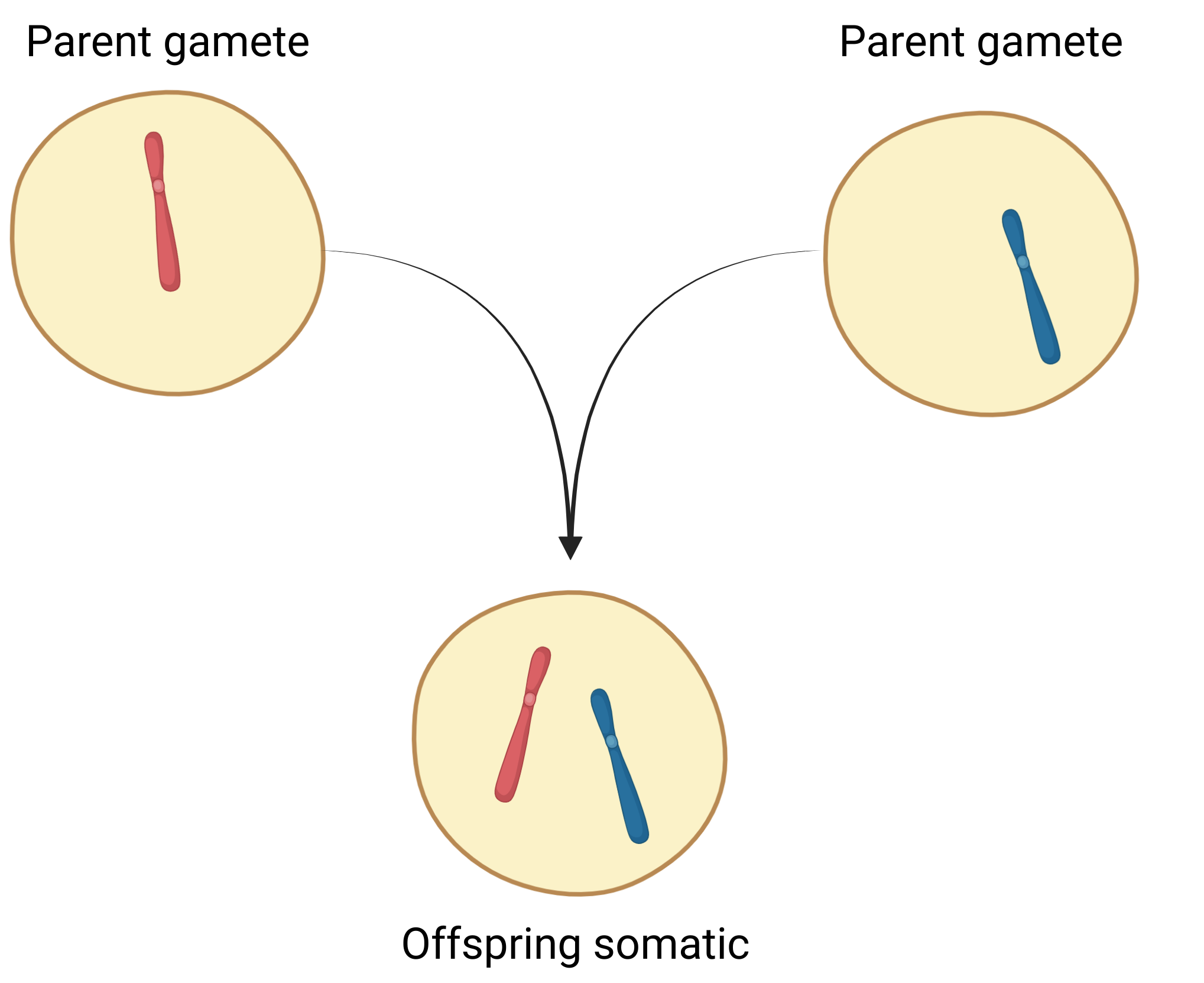
- In sexual reproduction, the organism creates a gamete reproductive cell. A gamete from a male parent and a gamete from a female parent combine, creating a child organism.
- Meiosis is the process of a regular cell dividing into and creating gametes.
DNA
DNA:
- DNA is a molecule within the nucleus of a cell which contains all the instructions for the cell.
- A gene is a unit of DNA with a specific instruction set.
- The genome is the totality of an organism’s DNA.
DNA Diversity:
- Every organism has a unique DNA.
- While organisms of a species have mostly similar DNA, there are slight variations from organism to organism, resulting in each organism’s individual characteristics.
- The main focus of cellular division is ensuring that each daughter cell receives properly copied DNA.
DNA Organization
Chromatin: DNA is packaged into chromatin
- The DNA double-helix molecule wraps around histone protein.
- Each unit of DNA-histone is a nucleosome.
- Nucleosomes coil into chromatin.
Chromosome: The chromatin forms chromosome structures
- In prokaryotes, the entire DNA is in a single circular chromosome.
- In eukaryotes, the DNA is divided between multiple linear chromosomes.
- During most of the cellular cycle, the chromosome chromatin is spread out, and chromosomes are not visible.
- Prior to cell division, the chromosome chromatin condenses into a defined chromosome structure, and the chromosomes are visible.
Haploid, Diploid
Haploid:
- Haploid cells are cells with a single set of chromosomes.
Diploid:
- Many organisms have diploid cells; cells with two sets of chromosomes.
- In diploid cells, one set of chromosomes comes from the male parent, the other from the female parent.
Somatic, Gametes
Somatic:
- In humans, all cells, aside from reproductive cells, are diploid. These are called somatic cells.
Gametes:
- The reproductive cells, however, are haploid cells, called gametes. This includes the male’s sperm cells and female’s egg cells.
- During meiosis, a somatic cell, with two sets of chromosomes, divides into gamete cells.
- Each gamete cell is haploid, and has only one set of chromosomes, randomly selected from between the original somatic cell’s two sets.
- A child organism is created from the combination of a male parent gamete and a female parent gamete.
- This creates a child somatic cell, with two sets of chromosomes, one from each parent gamete.
Human Chromosomes
Human Chromosome Count:
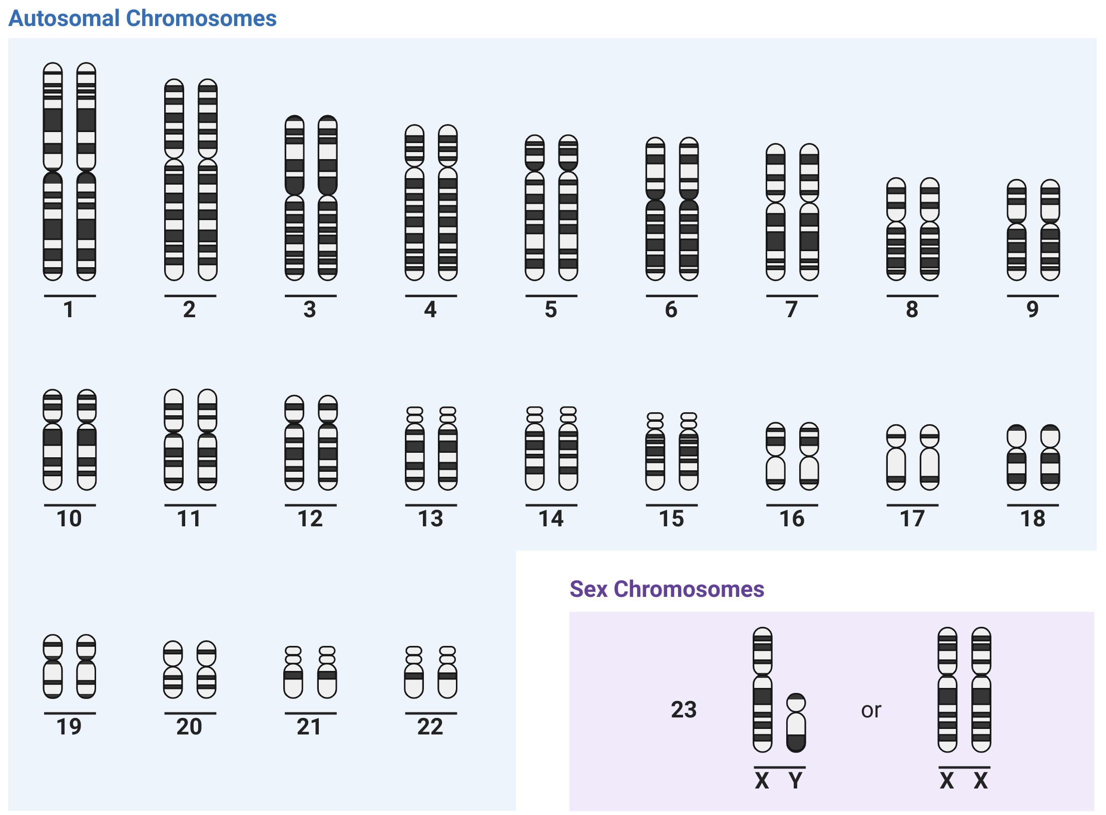
- The human genome is divided into 23 chromosomes.
- In diploid somatic cells, which have 2 sets of chromosomes, this totals 46 chromosomes.
- This is referred to as 2n; n being the number of chromosomes per DNA set, 2 being the number of each chromosome. In humans, n is 23; 2n is 46.
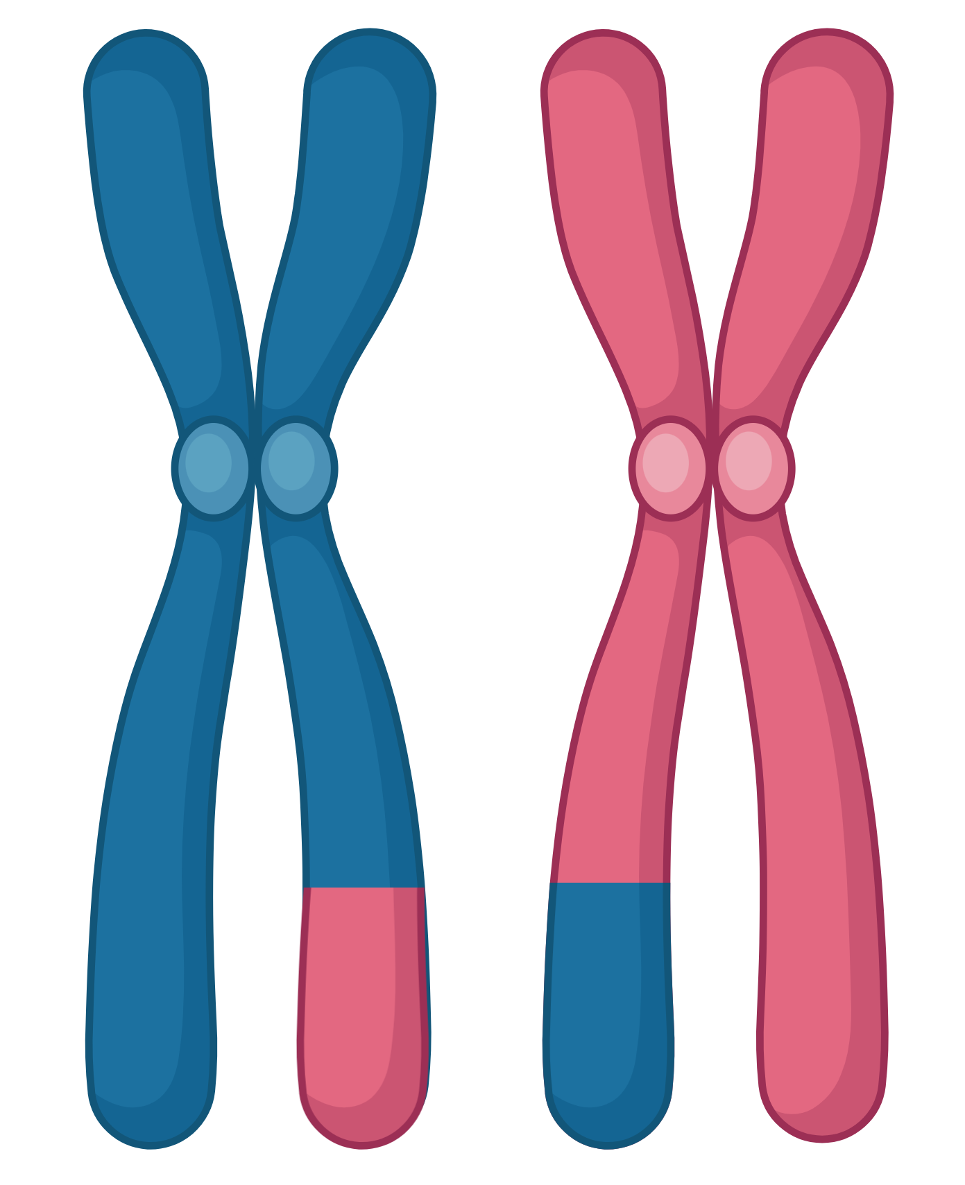
- Each pair of matching chromosomes is a homologous pair; the two chromosomes are homologues.
Autosomes, Sex Chromosomes
- The first 22 pairs of chromosomes, called autosomes, are nearly identical.
- The 23rd chromosome pair, the sex chromosomes, determines the organism’s sex, and may be different:
- If both chromosomes are ‘x’ chromosomes, the organism is female.
- If one is ‘x’ and one is ‘y’, the organism is male.
Cell Division Overview
Cell Division Prep
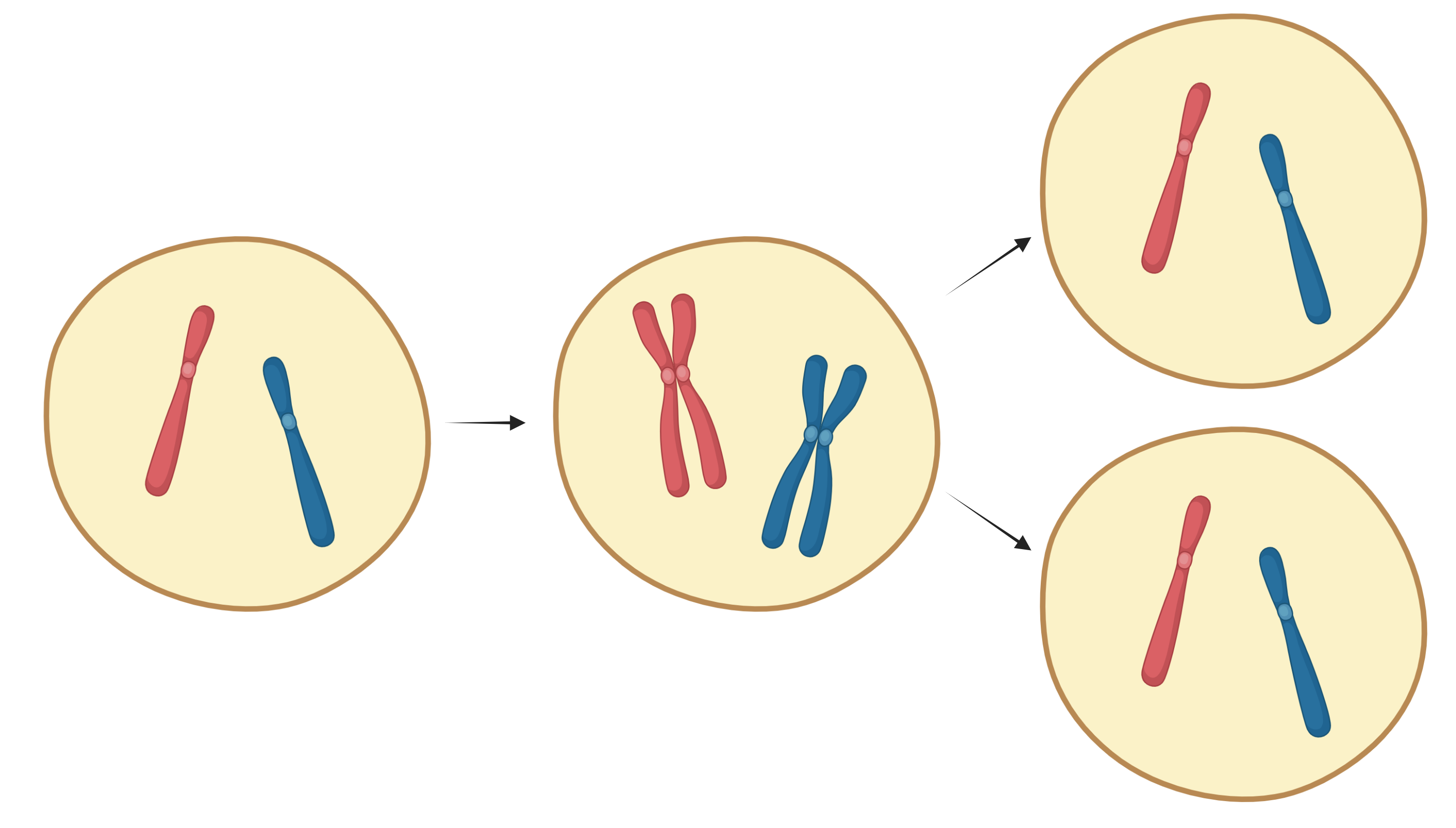
- Initially, each of the 46 chromosomes are an individual chromatid.
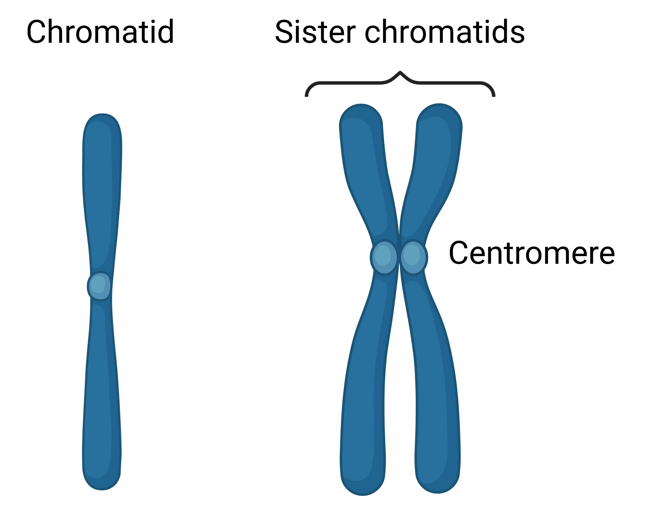
- Prior to cell division, each chromatid duplicates, creating 46 pairs of identical sister chromatids.
Mitosis
- During mitosis, the sister chromatids are split into the two daughter cells, resulting in two daughter cells with identical DNA.
Meiosis:
- During meiosis, after the chromatids duplicate and create sister chromatids, the homologous pairs are divided into two haploid cells, each with 23 chromosomes, each of those 23 chromosomes still containing two sister chromatids. This is the first step of meiosis, named Meiosis I.
- In Meiosis II, each haploid cell’s sister chromatids split into separate cells, much like mitosis.
- In total, meiosis results in 4 daughter haploid cells.
- Meiosis takes place in the female ovaries and male testes
Mitosis
Cell Cycle
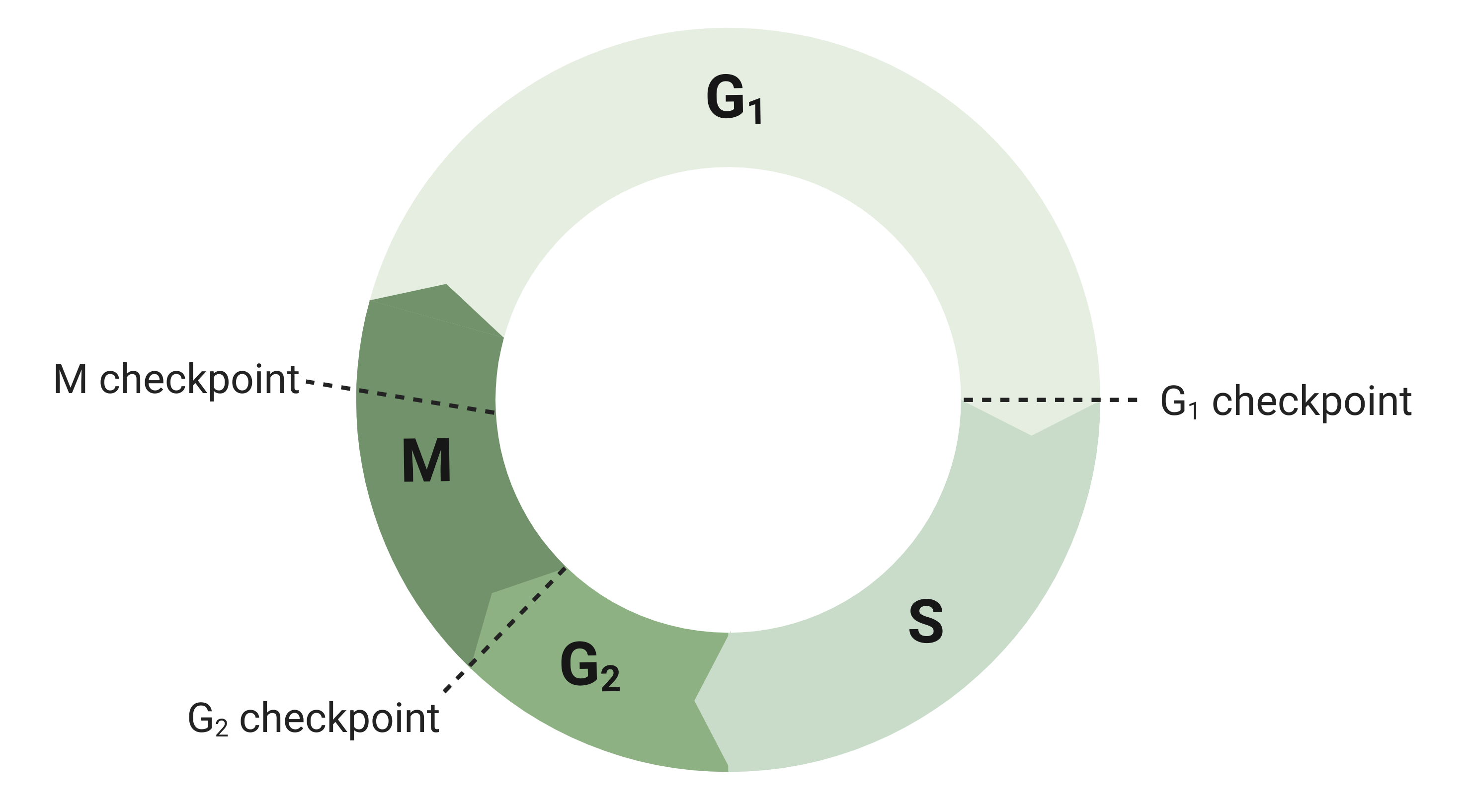
The cell cycle is the cycle a cell takes from its birth to when it divides and forms new cells.
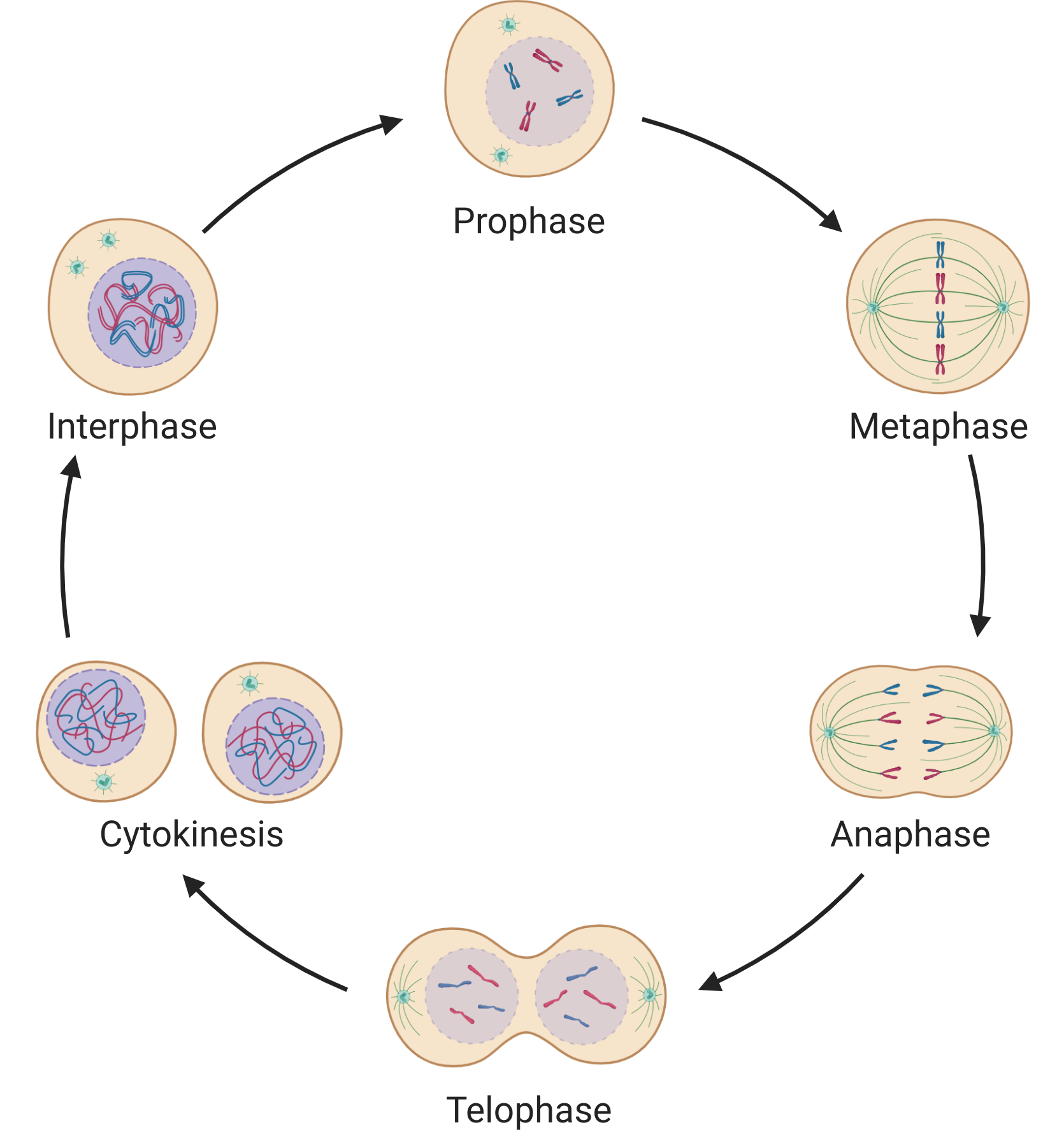
The cell cycle contains two phases: interphase and the mitotic phase.
Interphase
Interphase:
- The majority of the cell cycle is interphase.
- During interphase, the cell prepares for division.
- Interphase has three phases - G1, S, and G2:
- $G_1$ - Gap 1: The cell grows and prepares for DNA duplication
- $S$ - Synthesis: The DNA is duplicated
- Each chromatid is duplicated, forming pairs of sister chromatids
- The sister chromatids of each pair are joined at their centromeric region
- The centrosomes - and centrioles where applicable - are also duplicated
- $G_2$ - Gap 2: The cell grows further and prepares for the mitotic phase
Mitotic Phase
Mitotic Phase:
- During the mitotic phase, the sister chromatids are divided into two separate nuclei
- The mitotic phase has numerous subphases:
Steps of the Mitotic Phase
Prophase:
- The chromosomes condense and become visible
- The centrosomes - and centriole where applicable - begin moving to opposite poles of the cell and begin forming the mitotic spindle
- The centrosomes are anchored at the poles by astral microtubules
- The nuclear envelope breaks down down
Metaphase:
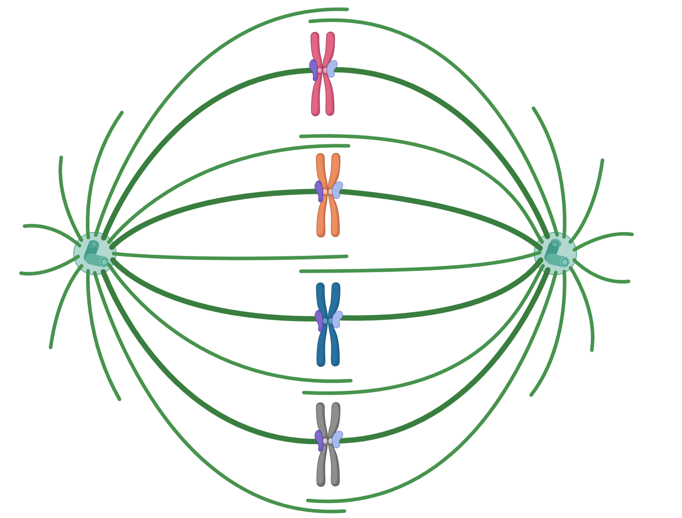
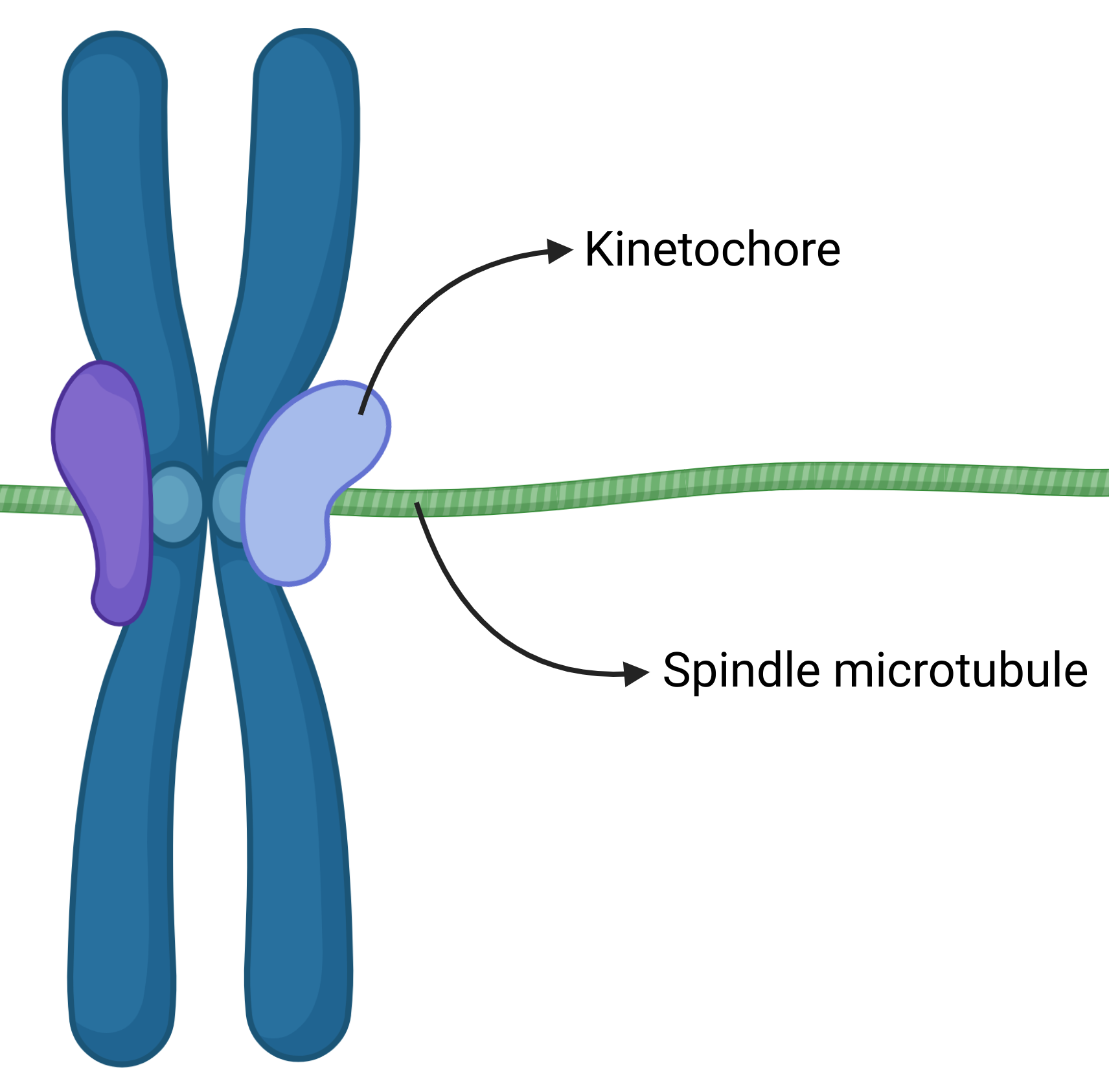
- Kinetochore is formed on the centromeric region of each chromatid
- The mitotic spindle microtubules extend and attach to the kinetochore
- The kinetochore and spindle microtubules perfectly line up the chromosomes at the metaphase plate in the center of the cell
Anaphase:
- The sister chromatids are split
- The split chromatids are pulled to their respective centrosomes on opposite sides of the cell
Telophase:
- The chromosomes are fully segregated to opposing poles; two new nuclear membranes form; the chromosomes decondense
- The mitotic spindle microtubules disassemble
Cytokinesis
Cytokinesis:
- Once the cell cycle is finished, and the two nuclei are complete, the actual cell divides, forming the two new daughter cells
Animal cell cytokinesis:
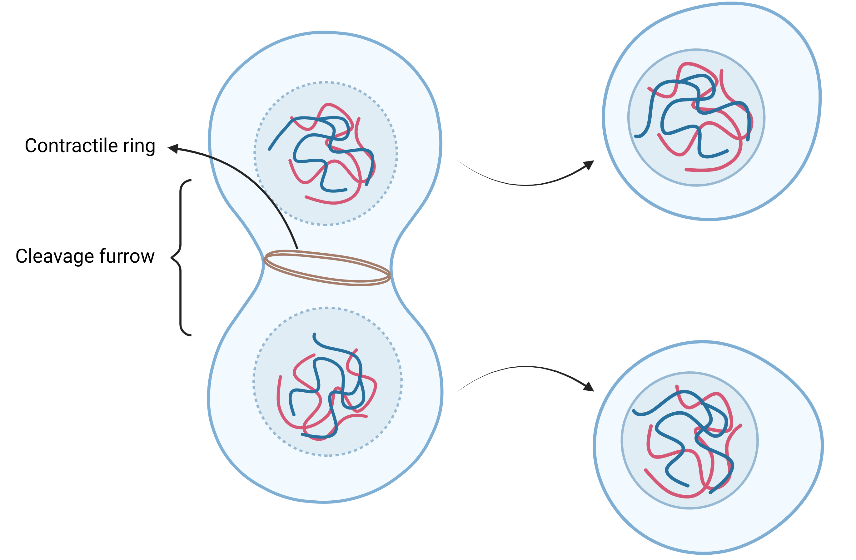
- In animals cells, cytokinesis takes place via a cleavage furrow
- Actin filaments form a contractile ring around the interior of the center of the cell
- The contractile ring constricts, pinching in the cell membrane, causing the opposing sides of the cell membrane to eventually meet in the middle, fuse together, and form two separate cells
Plant cell cytokinesis:
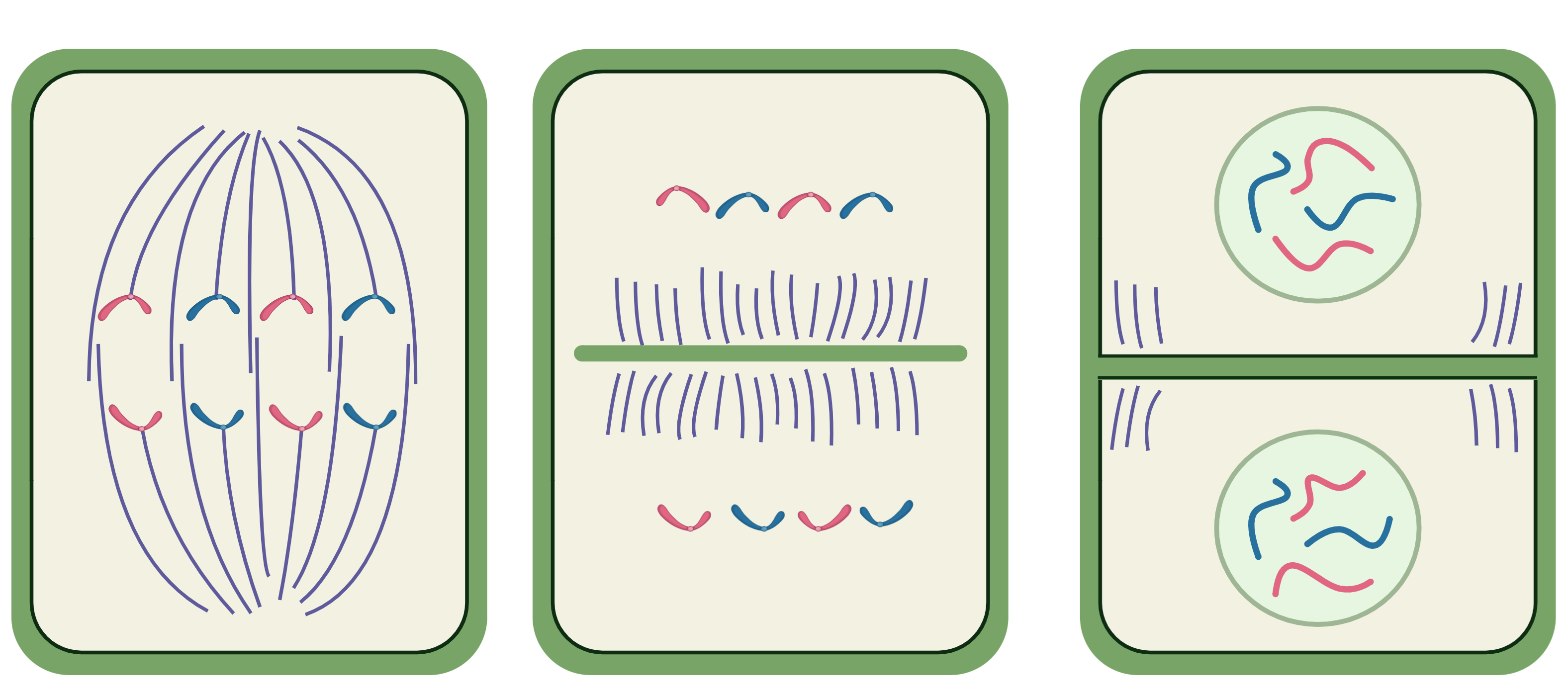
- Plant cells cannot divide via a cleavage furrow, pinching in their sides, due to their rigid cell wall
- Instead, vesicles from the Golgi apparatus form a cell plate in the center of the cell, eventually constructing an entirely new membrane and wall, dividing the cell in two
Cell Cycle Checkpoints
Cell Cycle Checkpoints:
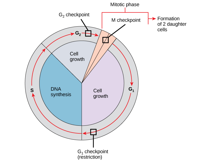
- The cell cycle has three checkpoints.
- When a checkpoint is met, the cycle pauses, and only resumes one it is ensured that certain conditions are met.
- Checkpoints also prevent the division of damaged cells
$G_1$ Checkpoint:
- The first checkpoint is the G1 checkpoint
- The G1 checkpoint takes place during interphase, prior to the S phase
- The G1 checkpoint checks that the cell is able to duplicate the DNA and proceed with cell division
$G_2$ Checkpoint:
- The second checkpoint is the G2 checkpoint
- The G2 checkpoint takes place at the end of interphase, prior to the mitotic phase
- The G2 checkpoints checks that the DNA was properly duplicated
M Checkpoint:
- The final checkpoint is the M checkpoint
- The M checkpoint takes place during the mitotic phase, at metaphase
- The M checkpoint checks that all the chromosomes are correctly lined up at the center of the cell and their kinetochore is properly attached to the spindle fibers
Failed Checkpoints:
- A characteristic of cancerous cells is unregulated growth and division
- This generally entails a cell checkpoint failing
Meiosis
Meiosis Overview
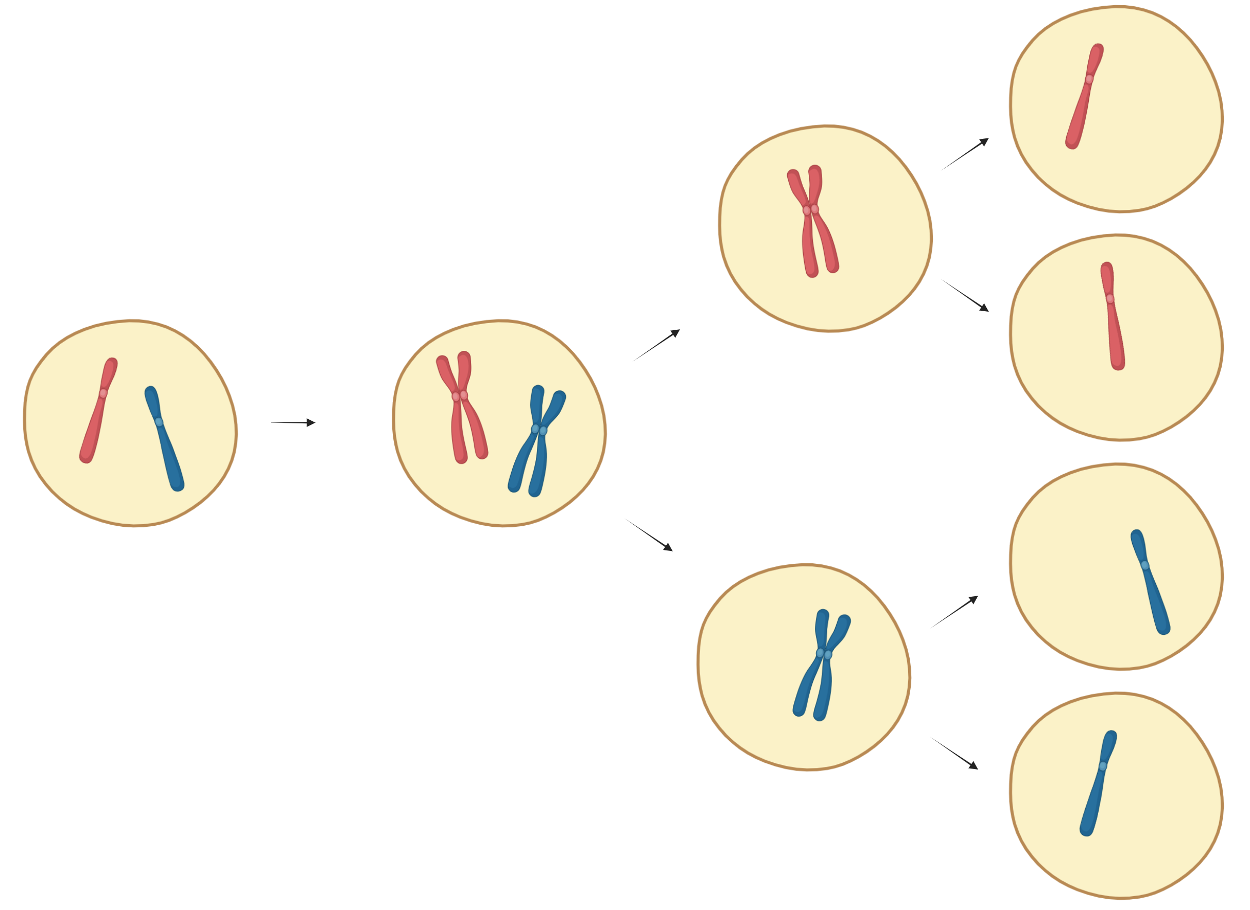
During meiosis, a somatic cell divides into haploid gamete cells.
Meiosis has two stages:
- Meiosis I: The homologous pairs separate into two haploid cells
- Meiosis II: The sister chromatids in the haploid cells split into two separate cells
- In total, meiosis produces four daughter cells
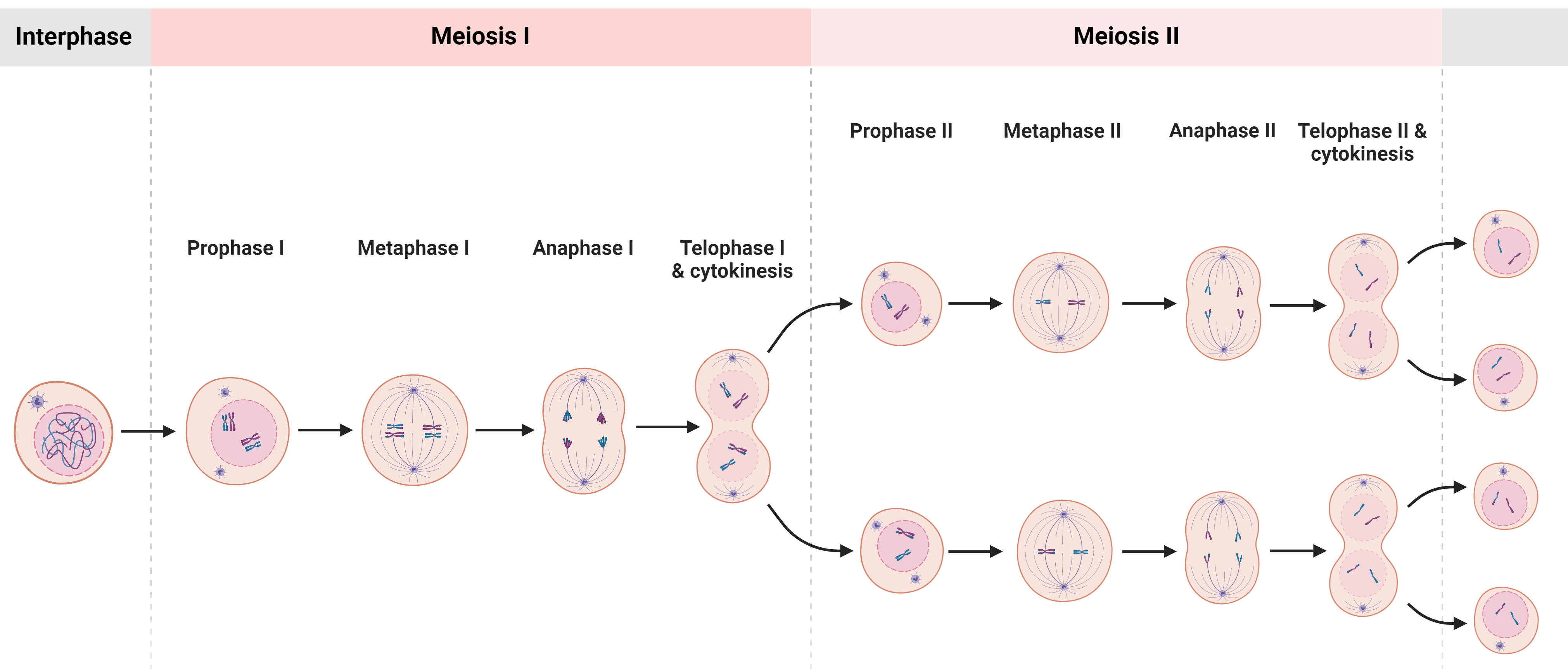
Interphase
Prior to meiosis, the cell undergoes interphase, similar to mitosis.
During interphase, the DNA and chromosomes duplicate.
Meiosis I
Prophase I:
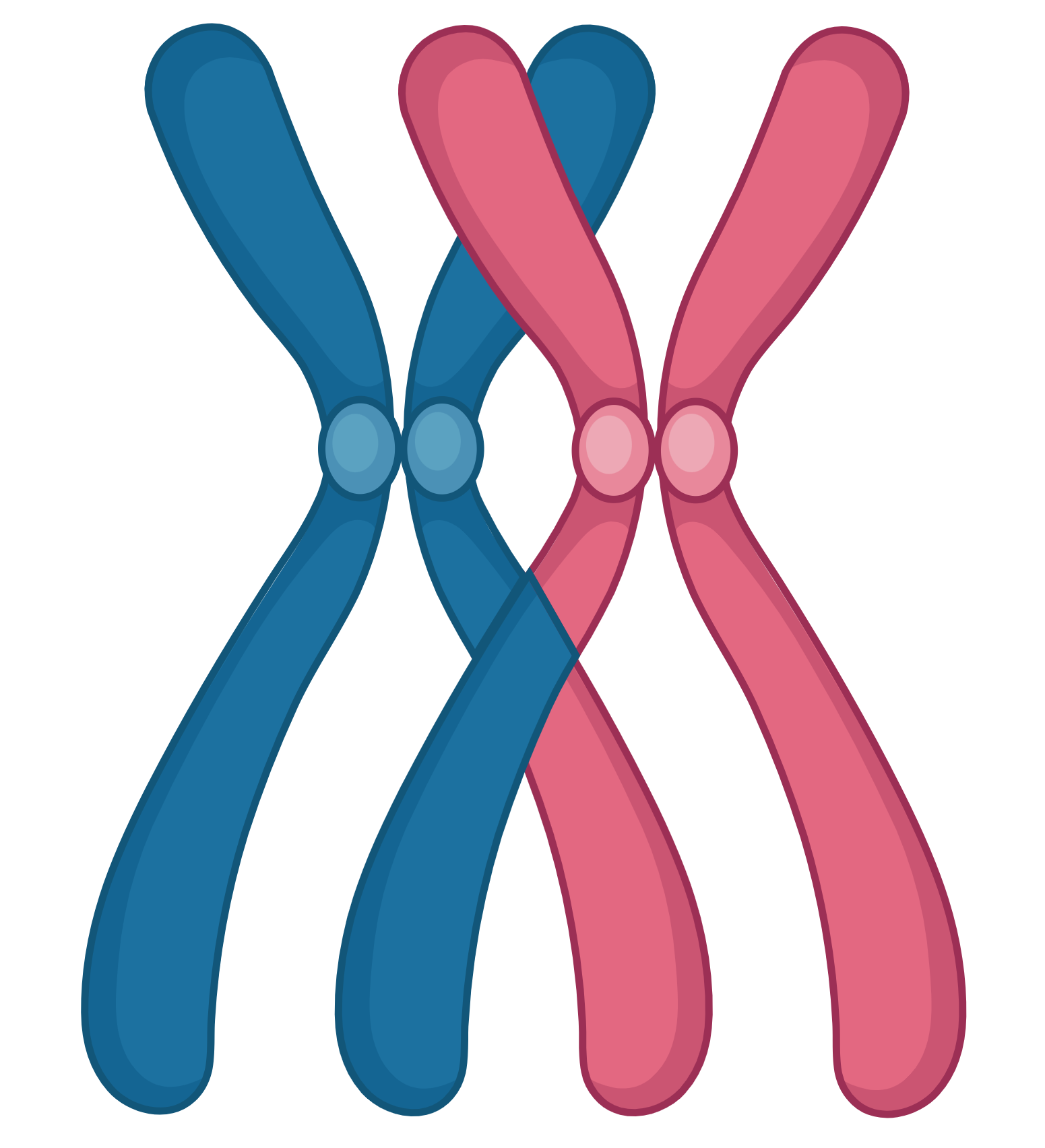
- During prophase I, the homologues align and attach to each other, forming tetrads
- Some genetic information is exchanged between the homologues, increasing the resultant child cell’s diversity
- Spindle microtubules attach to each homologue’s kinetochore
Metaphase I:
- The tetrads arrange at the mitotic plate
Anaphase I:
- The homologue chromosomes are pulled to opposite centrosome poles
Telophase I, Cytokinesis I
Meiosis II
During meiosis II, the haploid cells’ sister chromatids divide, similarly to mitosis.
Meiosis II includes:
Prophase II, Prometaphase II, Metaphase II, Anaphase II, Telophase II, Cytokinesis II
Schematics
Overview
-
DNA: see chapter 3 and 4
-
Cell cycle, checkpoints
-
Gamete, somatic
-
Human genome
Mitosis
-
Mitosis overview
-
Interphase synthesis
-
Mitotic phases
-
Metaphase, kinetochore
-
Kinetochore
-
Animal cytokinesis
-
Plant cytokinesis
Meiosis
-
Meiosis overview
-
Meiosis phases
-
Tetrad
-
Chromosomes with exchanged material